What is High-Risk Proliferative Diabetic Retinopathy?
High-Risk Proliferative Diabetic Retinopathy (PDR) refers to a state that puts patients at severe risk of developing diabetic retinopathy due to which they may develop complete or partial vision loss. We have covered in-depth how to prevent diabetic retinopathy here.
Hence, there is a need to be aware of what high-risk PDR means to be able to avoid these complications. Here is a brief discussion about what PDR is and the key features of this condition.
What is High-Risk Proliferative Diabetic Retinopathy (PDR)?
High-Risk Proliferative Diabetic Retinopathy refers to a state that puts patients at a greater risk of developing Diabetic Retinopathy due to which they may develop complete or partial vision loss. Proliferative diabetic retinopathy can be categorized into early proliferative diabetic retinopathy or high-risk proliferative diabetic retinopathy.
The early proliferative diabetic retinopathy is characterized by the formation of new blood vessels in the eyes, though it does not meet the criteria for the diagnosis of high-risk PDR. However, if not detected early or managed properly, early proliferative diabetic retinopathy may progress resulting in high-risk proliferative diabetic retinopathy.
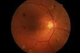
In patients with high-risk PDR, the neovascularization of the disc (NVD) affects at least one-third of the disc area (DA).
Neovascularization refers to the process of formation of new blood vessels in any tissue of the body. In patients with diabetes, neovascularization occurs as a response of the body to protect the eyesight. This occurs when the body perceives that there is damage to the blood vessels supplying oxygen and vital nutrients to the eyes and tries to compensate for it by triggering the formation of new blood vessels.
Hence, neovascularization is considered one of the defense mechanisms of the body that is aimed at ensuring the vital structures are not deprived of the blood supply.
However, in patients with diabetes, neovascularization can result in the formation of new blood vessels that are not strong and hence, vulnerable to rupture. So, these newly formed blood vessels in the eyes may burst resulting in hemorrhage within the vitreous humor of the eye.
These abnormal changes in the eyes are responsible for the gradual loss of eyesight in patients with diabetes.
In some diabetic patients, neovascularization may affect more than half of the disc area resulting in considerable loss of vision.
The rapid progress of NVD can lead to pre-retinal or vitreous hemorrhage. If not detected and treated properly, these changes can become worse resulting in considerable damage to the vital parts of the eyes due to which patients may develop vision loss.
Hence, it is important to be aware of the initial warning signs of proliferative diabetic retinopathy so that it can be diagnosed at an early stage.

Why is Proliferative Diabetic Retinopathy Classified as High Risk?
PDR is classified as a high-risk form of retinopathy as the development of this condition is marked by a significant loss of vision.
In some cases, a combination of two mechanisms including traction and rhegmatogenous retinal detachments may occur in patients with PDR putting them at a much greater risk of vision loss.
Macular edema caused due to these abnormal changes in the retina is one of the leading causes of visual impairment, especially in patients with uncontrolled diabetes. It may occur due to functional damage or necrosis of the small capillaries in the retina.
In patients with proliferative diabetic retinopathy, edema may also be caused due to retinal traction when the retina has been elevated away from the epithelial tissues.
These are some reasons why proliferative diabetic retinopathy is classified as ‘high risk’. This also emphasizes the need to be watchful about the early signs of this condition in order to protect your eyesight.
Read on to learn more about the key characteristics of proliferative diabetic retinopathy that could help in the early diagnosis of this condition.
What Are the Key Characteristics of Proliferative Diabetic Retinopathy?
Neovascularization
It is the hallmark sign of PDR. It usually occurs near the disc-like structure in the eyes called the optic disc resulting in the state called the neovascularization of the disc (NVD).
Neovascularization may also occur within three-disc diameters of the major blood vessels of the retina in which case it is referred to as the neovascularization elsewhere (NVE).
Preretinal Hemorrhages
It is a sign that appears as small pockets of blood present in the potential spaces between the posterior hyaloid face and the retina. It may occur due to the pooling of the blood usually in the shape of a boat.
Hemorrhage into the vitreous
The newly formed blood vessels in the eye are weak and vulnerable to rupture due to which patients may develop hemorrhage in the vitreous humor. It often appears as diffuse hazing or clumps of clots in the gel.
Fibrovascular tissue proliferation
The fibrovascular proliferation of the tissue is usually associated with neovascular complexes. It may appear avascular if the blood vessels have become regressed.
Traction retinal detachments
It usually appears tented up, concave, and immobile, as compared to the rhegmatogenous retinal detachments that look bullous, convex, and mobile. A combination of both these mechanisms is not uncommon in patients with diabetes and high-risk proliferative diabetic retinopathy.
Macular edema
Macular edema is one of the leading causes of visual impairment in patients with diabetes. It often occurs due to damage to the retinal capillaries. Edema also may be caused due to retinal traction when the retina has been sufficiently pushed away from the epithelial tissues.
Know your risk of diabetic eye disease
Click here to download the RetinaRisk app now!


How Does RetinaRisk App And API Help To Detect High-Risk Proliferative Diabetic Retinopathy?
The RetinaRisk App and API are designed by a team of visionary doctors with an aim to provide personalized care to patients based on their individual risk assessment.
The clinically validated RetinaRisk app includes an advanced algorithm that can empower you to calculate your risk of developing any sight-threatening eye disease accurately allowing you to seek timely treatment for preserving your eyesight.
Conclusion
RetinaRisk App can allow you to assess your eye health and vision regularly and help you in the early diagnosis of high-risk PDR. This would protect you against the more serious complications including blindness.


